
Countstar® Rigel S5
Advanced Image Cytometer

The Countstar Rigel S5 system increases the PE channel on the basis of Rigel S3, and can carry out multi-color fluorescence experiments in addition to rapid cell viability and concentration measurement, including cell apoptosis (Annexin V/PI/DAPI), cell transfection (GFP/YFP/RFP), CD marker experiment (DAPI/FITC/PE), and cell killing (Hoechst T33342/Calcein-AM/PI) and so on.
Excitation light: 375nm, 480nm, 525nm
Emission filter:460nm, 535nm, 580nm, 600LP
Key Benefits
All in one design- Integrated Computer/Touch scree
Automatic Detection 5 Samples in one single test sequence.
User Friendly Cell Analysis Protocols
Unrivaled "Fixed focus" patent, no manual focusing.
High Stability and Actuary
Powerful and user-friendly Data Management
GMP and 21 CFR part 11 ready
FCS Express Software
Product feature
SmartThe BioApps of the Countstar Rigel S3 System simplify routine cell lab tasks while providing high quality scientific data.
Countstar S3 comes with three BioApps to simplify and automate the routine cell count, viability and cytotoxicity tasks.
 AO/PI Viability Protocol: Run two fluorescence color assays in disposable consumable to determine the percentages of live, dead cells and concentration in the presents of debris and unwanted nonnucleated cell types including red blood cells.
 Affinity of Antibody: This assay run as Green color assays in FITC binding antibody to determine the affinity of the biosimilar drug.
 Surface Marker Assay: Quantify specific cell populations based on surface marker expression (CD45+ CD4+, CD8 + MSC, CD56+ NK cells, etc.)
 Cell Cycle: Propidium iodide (PI) is a nuclear staining dye that is frequently applied in measuring cell cycle. The protocol determining cellular DNA content in cell cycle analysis
 Cell Apoptosis: Cell Apoptosis assay is a type used for determining the apoptosis percentage of cells by Annexin V-FITC/7-ADD staining method.
 GFP transfection: The green fluorescent protein (GFP) exhibits bright green fluorescence when exposed to light in the blue to ultraviolet range. This protocol can analyze counting and percentage of GFP.
 Celling Killing: Run three fluorescence color assays in disposable consumable to determine the CAR T/NK-Mediated Cytotoxicity using Tracer and Viability Dyes
 Trypan blue Protocols: Obtain cell count, viability and concentration estimations based on trypan blue staining using a disposable consumable.
Three step process

Reproducible and Accurate

Touch Screen with Flexible Experiment Assay Management System

FCS Express Software

GMP ready

Applications
Dual-Fluorescence Viability


T/NK cell mediated cytotoxicity


Cell Apoptosis

Cell Cycle
CountstarRigel S5 enables users to get the result of a cell cycle quickly and accurately.CounstarRigel S5 can analyzeproportionof cell indifferentphase of the cell cycle.

Surface Maker Analysis
A lymphocyte subsets analysis is a typical experiment performed incell relatedresearch fields and various diseases diagnosis. Countstar® Rigel S5 offers a faster and easier way to make immune cell typing more efficient. With visible cell images and powerful data analysis

Affinity of antibody
Detect affinity of antibody in cell level is an important indicator of monoclonal antibody detection using immunofluorescence method.TheCountstar® Rigel S5 offers a rapid, direct and reliable evaluating method for the affinity of antibody detection in antibody drug screening

GFP Transfection Efficiency
In cell and molecular biology, the GFP gene is frequently used as a reporter of expression. Currently, scientists are commonly using the fluorescent microscopes or flow cytometers to analyze the transfection efficiency of mammalian cells. But Flow cytometry requires a high-qualified and experienced operator. WhileCountstarRigel S5 enables users to get the result of a transfection efficiency assay quickly and accurately.

Advanced Image Cytometer

- PBMC and Primary Cell Counting
- Cell Apoptosis
- Cell Cycle
- Surface Marker Analysis
- GFP Transfection Efficiency
- FCS Express Software
The Countstar Rigel S5 system increases the PE channel on the basis of Rigel S3, and can carry out multi-color fluorescence experiments in addition to rapid cell viability and concentration measurement, including cell apoptosis (Annexin V/PI/DAPI), cell transfection (GFP/YFP/RFP), CD marker experiment (DAPI/FITC/PE), and cell killing (Hoechst T33342/Calcein-AM/PI) and so on.
Excitation light: 375nm, 480nm, 525nm
Emission filter:460nm, 535nm, 580nm, 600LP
All in one design- Integrated Computer/Touch scree
Automatic Detection 5 Samples in one single test sequence.
User Friendly Cell Analysis Protocols
Unrivaled "Fixed focus" patent, no manual focusing.
High Stability and Actuary
Powerful and user-friendly Data Management
GMP and 21 CFR part 11 ready
FCS Express Software
SmartThe BioApps of the Countstar Rigel S3 System simplify routine cell lab tasks while providing high quality scientific data.
Countstar S3 comes with three BioApps to simplify and automate the routine cell count, viability and cytotoxicity tasks.
AO/PI Viability Protocol: Run two fluorescence color assays in disposable consumable to determine the percentages of live, dead cells and concentration in the presents of debris and unwanted nonnucleated cell types including red blood cells.Affinity of Antibody: This assay run as Green color assays in FITC binding antibody to determine the affinity of the biosimilar drug.Surface Marker Assay: Quantify specific cell populations based on surface marker expression (CD45+ CD4+, CD8 + MSC, CD56+ NK cells, etc.)Cell Cycle: Propidium iodide (PI) is a nuclear staining dye that is frequently applied in measuring cell cycle. The protocol determining cellular DNA content in cell cycle analysisCell Apoptosis: Cell Apoptosis assay is a type used for determining the apoptosis percentage of cells by Annexin V-FITC/7-ADD staining method.GFP transfection: The green fluorescent protein (GFP) exhibits bright green fluorescence when exposed to light in the blue to ultraviolet range. This protocol can analyze counting and percentage of GFP.Celling Killing: Run three fluorescence color assays in disposable consumable to determine the CAR T/NK-Mediated Cytotoxicity using Tracer and Viability DyesTrypan blue Protocols: Obtain cell count, viability and concentration estimations based on trypan blue staining using a disposable consumable.Three step process
The Countstar S3 System is designed to take you from sample to result in one go, simplifying your work and allowing for increased productivity and efficiency.

Reproducible and Accurate
Countstar S3 system can automatic detect 5 samples at one time, it will be more efficiency and affordable, with integrated patent “fixed focus”, the results would be more stable and reliable.

Touch Screen with Flexible Experiment Assay Management System
“App-like" interface. Allow to add multiple Experiment Assays for different operators

FCS Express Software
The optional De NovoTM FCS Express software makes graphs touchable and customizable fluorescence channel boost your experiment reach. Countstar Rigel S5 together with FCS Express is able to analyze for the cell apoptosis, cell cycle, transfection, affinity of antibody, CD marker and etc.

GMP ready
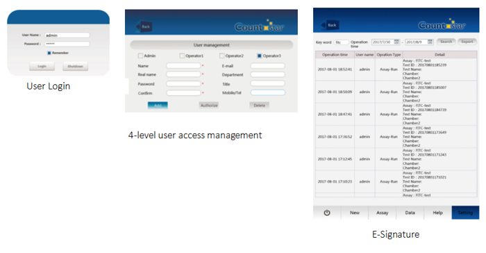
The Countstar S5 system is designed to meet the modern pharmaceutical requirements. The software complies with 21 CFR part 11. Key features include control of user access and data export, and secure audit trail. At the same time, we also supply IQ/OQ service and PQ support to help customer build a reliable system.
Specifications
| Technical Specifications | |
| Model: | Countstar Rigel S5 |
| Diameter range: | 3μm ~ 180μm |
| Concentration range: | 1×10⁴ ~ 3×10⁷/mL |
| Objective magnification: | 5x |
| Imaging element: | 1.4 megapixel ,CCD camera |
| Excitation Light | 375nm, 480nm, 525nm |
| Emission Filter | 460nm, 535nm, 580nm, 600LP |
| USB | 1×USB 3.0 1×USB 2.0 |
| Storage: | 500GB |
| Power supply: | 110–230 V/AC, 50/60Hz |
| Screen: | 10.4 inch touchscreen |
| Weight: | 13kg (28lb) |
| Size (W X D X H): | Machine: 254×303×453mm Package size: 430×370×610mm |
| Operating temperature: | 10℃ ~ 40℃ |
| Working humidity: | 20% ~ 80% |
Dual-Fluorescence Viability
Acridine orange (AO) and Propidium iodide (PI) are nuclear nucleic acid binding dyes. The analysis excludes cell fragments, debris and artifacts particles as well as undersized events such as platelets, giving a highly accurate result. In conclusion, theCountstarsystem can be used for every step of the cell manufacturing process.


By labeling the target tumor cells with CFSE ortransfectwith GFP. Hoechst 33342 is used for stain all cells (both T cells and tumor cells), alternatively, target tumor cells can be stained with CFSE, PI is used forstainthe dead cells (both T cells and tumor cells). This staining strategy allows for the discriminate of different cells.


Cell Apoptosis
The different phases of apoptosis are segmented into an early, middle and late stage. Specified indicators can document the single stages of apoptosis. The Countstar® Rigel S5 offers a visualized image-based solution and analyzes all of these steps with pre-loaded protocols in depth.

CountstarRigel S5 enables users to get the result of a cell cycle quickly and accurately.CounstarRigel S5 can analyzeproportionof cell indifferentphase of the cell cycle.

A lymphocyte subsets analysis is a typical experiment performed incell relatedresearch fields and various diseases diagnosis. Countstar® Rigel S5 offers a faster and easier way to make immune cell typing more efficient. With visible cell images and powerful data analysis

Detect affinity of antibody in cell level is an important indicator of monoclonal antibody detection using immunofluorescence method.TheCountstar® Rigel S5 offers a rapid, direct and reliable evaluating method for the affinity of antibody detection in antibody drug screening

In cell and molecular biology, the GFP gene is frequently used as a reporter of expression. Currently, scientists are commonly using the fluorescent microscopes or flow cytometers to analyze the transfection efficiency of mammalian cells. But Flow cytometry requires a high-qualified and experienced operator. WhileCountstarRigel S5 enables users to get the result of a transfection efficiency assay quickly and accurately.

评论
发表评论