Countstar® BioTech
Automated Cell Counter
Accurate cell analysis for:
Product Overview
The Countstar® is designed based on the classic trypan blue staining for cell viability principle, integrating the advanced “fix focus” optical imaging work bench and the most advanced cell recognition technologies and software algorithms. Countstar BioTech not only provides the cell concentration and viability, but also provides the average diameter and average circularity, and showing the diameter range and aggregation of cells in histograms etc.
Key Benefits
Product feature
Easy and fast:
Cost-efficient Consumables:
Loaded 5 sample in one slide Individual Package
Imaging Technology:
Accuracy and Reliability:
Guaranteed by “Fixed Focus” technology, high resolution imaging, larger observation area and advanced algorithms.
Countstar VS Hemocytometer
Powerful data analysis and management System:
1. Intelligence search, Coded lock, security user login and sustainable data management etc., to keep the cell quality and data safety.
2. Countstar software system provides cultivation time chart (CTC), overlay analysis and other statistical and analytical functions
3. Diversity of Data Formats: PDF, EXCEL, JPEG;Automated PDF reports
4. Sustainable Data Management
User, Time, Parameter, Result……
1. Trypan blue cell counting and viability
2. Aggregated Cell Analysis
3. Cell Size Analysis
4. Overlay Analysis of the Cell Growth Curve
Automated Cell Counter
Accurate cell analysis for:
- Trypan blue Cell Counting
- Aggregated Cell Analysis
- Cell Diameter Analysis
- Cultivation time chart (CTC)
The Countstar® is designed based on the classic trypan blue staining for cell viability principle, integrating the advanced “fix focus” optical imaging work bench and the most advanced cell recognition technologies and software algorithms. Countstar BioTech not only provides the cell concentration and viability, but also provides the average diameter and average circularity, and showing the diameter range and aggregation of cells in histograms etc.
- “Fixed Focus”– No manual focus adjustment needed
- Fast testing time – less than 20s
- Data easily comparable with a hemocytometer
- Maximum loaded 5 samples in one slide
- Accurate and high reproducibility result – Intra-assay & Inter-assay CV <5%
- Maintenance and service free
- Powerful software and data management System
- IQ/OQ validation and PQ support
Easy and fast:
Within 20 seconds through three steps with one button
Loaded 5 sample in one slide Individual Package
Imaging Technology:
5-megapixel color imaging and a wide sample collection range ensures clear and accurate visualization
Accuracy and Reliability:
Guaranteed by “Fixed Focus” technology, high resolution imaging, larger observation area and advanced algorithms.
Countstar VS Hemocytometer
1. Intelligence search, Coded lock, security user login and sustainable data management etc., to keep the cell quality and data safety.
User, Time, Parameter, Result……
Specifications
Applications| Technical Specifications | |
| Model: | IC 1000 |
| Test Item: | Concentration, Viability, Diameter, aggregation etc. |
| Sample Density: | 1x10⁴ - 3x10⁷/ml |
| Sample Diameter: | 5-180μm |
| Imaging element: | 5 Megapixel,CMOS camera |
| Objective magnification: | 2.5 X |
| Sample Volume: | 20μL |
| Test Time: | <20s |
| Output: | JPEG/PDF/EXCE |
1. Trypan blue cell counting and viability
Countstar is applicable to cells with diameter between 5-180um, like mammalian cell, insect cell, and some planktons.
Some primary cells or subculture cells are prone to aggregate when poor culture state or excessive digestion, thus causing great difficulty in the counting of cells. With the Aggregation Calibration Function, Countstar® can realize a stimulation calculation of aggregations to ensure accurate cell counting and obtain the aggregation rate and the aggregation histogram, thus providing basis for experimenters to judge the state of cells.
Countstar can count the aggregated cells one by one.
Countstar can count the aggregated cells one by one.
 |
| HepG2 |
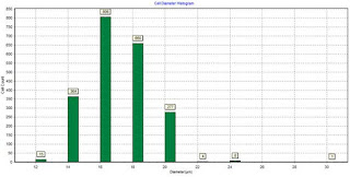
3. Cell Size Analysis
The change of cell size is a key feature and is commonly measured in cell research. Normally it will be measured in these experiments: cell transfection, drug test and cell activation assays.
4. Overlay Analysis of the Cell Growth Curve
In cell culture, the study of pharmacology and toxicology often requires the overlay analysis of multiple growth curves in order to find the optimal culture conditions or the best dosage. Countstar® can directly call up multiple growth curves for comparative analysis.


评论
发表评论